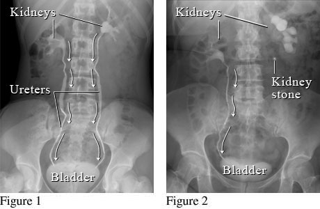
Courtesy of Intermountain Medical Imaging, Boise, Idaho.
These figures show an X-ray with contrast dye (intravenous pyelogram, or IVP) of the kidneys, ureters, and bladder. Figure 1 shows a normal flow from the kidneys, through the ureters, to the bladder (white arrows). Figure 2 shows a kidney stone blocking the normal flow of urine in the ureter on the right.
Current as of: December 19, 2022
Author: Healthwise Staff
Medical Review:E. Gregory Thompson MD - Internal Medicine & Brian D. O'Brien MD - Internal Medicine & Adam Husney MD - Family Medicine & Kathleen Romito MD - Family Medicine & Tushar J. Vachharajani MD, FASN, FACP - Nephrology